 What is ultrasound?
Ultrasound is a diagnostic test that is often performed after a physical exam, blood tests and/or radiographs have indicated a possible problem. An ultrasound machine uses sound waves to create images of internal structures such as the abdominal organs and heart. It can also be used to evaluate smaller structures such as the thyroid glands and tendons.
My pet just had x-rays. Why do we need an ultrasound?
An ultrasound is able to provide additional information that we are unable to obtain from traditional x-ray imaging. For example, abdominal organs and fluid have the same density. So, if the abdomen is full of fluid the abdominal organs cannot be differentiated on x-ray. However, with ultrasound they appear quite different, and even with the presence of fluid we are able to visualize the interior of specific organs such as the liver and heart.
On the other hand, ultrasound cannot be used to image bones or air-filled structures such as the lungs. The presence of air or gas can actually interfere with ultrasound imaging of different abdominal structures. This is partially why we require our patients be fasted prior to testing.
In a nutshell, ultrasound does not replace radiographs, but complements the information obtained with x-rays. It is common to do both x-rays and ultrasound to get a complete picture.
Is ultrasound imaging painful?
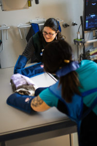
Not at all! In order to perform an ultrasound, the hair must be clipped from the area being evaluated. The pet is then placed in a padded trough (for abdominal ultrasound) in a darkened room. Just like in human medicine, ultrasound gel is used to obtain good contact between the skin and the probe while images are collected. Most patients do not require any sedation for an exam.
What is an ultrasound-guided fine needle aspirate?
This is a procedure used to obtain cells from areas that appear abnormal. Using the ultrasound, a very thin needle is guided into the area of interest to obtain cells. These cells are then placed on a slide and examined microscopically for evidence of cancer, infection, or inflammation. The discomfort experienced is expected to be similar to that of a blood draw and is well tolerated by most patients, although sedation is occasionally needed.
Is this procedure safe?
Ultrasound- guided fine needle aspiration is minimally invasive and can often be used to obtain a final diagnosis. It is especially helpful in cases where more invasive procedures, such as an exploratory surgery, are not an option. Although generally safe, potential risks associated with needle biopsy of deep structures can include bleeding or infection. It is a good idea to discuss this procedure with the veterinarian prior to scheduling an ultrasound.
Who should perform my pet’s ultrasound?
The collection and interpretation of ultrasound images is best done by a veterinarian with experience and special training in this imaging modality. Most of the ultrasound exams done at the Animal Clinic in Sussex are performed by Dr. Jennifer Cortright who has been using ultrasound for more than a decade and has completed a 2-year certification program in small animal ultrasound through the University of Illinois. Some examples of diagnoses made at ACS using ultrasound include tumors of the spleen, liver, stomach and thyroid, obstructed ureters, retained testicles, intestinal foreign bodies, and a ruptured Achilles tendon.
